Site hosted by Angelfire.com: Build your free website today!
« April 2019 »
S M T W T F S
1 2 3 4 5 6
7 8 9 10 11 12 13
14 15 16 17 18 19 20
21 22 23 24 25 26 27
28 29 30
Entries by Topic
All topics  «
Blog Tools
Edit your Blog
Build a Blog
RSS Feed
View Profile
You are not logged in. Log in
My expert blog 2072
Thursday, 4 April 2019
What I Dream Everyone Understood About CT Scan

A CT or CAT scan is a reduced name for electronic tomography. A CT scan takes pictures of the within the body. The pictures are more in-depth than a typical x-ray. During a CT scan of the chest, pictures are taken of random samples or slices of the thoracic structures in your body. The thoracic structures include your lungs, heart and the bones around these locations. When contrast is utilized during a CT scan of the chest thoracic structures are highlighted even more. CT scans can help determine a diagnosis early. Your doctor will use this info to identify the best treatment for you.

Using a CT Scan increases the detection of lung cancer at Stage 1 up to the rate of 85 percent. Lung cancer is often found when the tumor is reasonably large, which results in treatment that may need more extensive surgery, chemotherapy, and/or radiation therapy and less beneficial outcomes when compared to finding cancer previously. By utilizing low-dose spiral computed tomography (Spiral CT), an unique kind of X-ray imaging more sensitive than routine chest X-rays, we anticipate finding more lung cancer cases at an earlier stage of growth and enhancing survival rates. Another benefit to low-dose radiation imaging is that the quantity of radiation a person would get is https://en.search.wordpress.com/?src=organic&q=CT Scan substantially reduced as compared to that of a standard-dose diagnostic CT

. CT lung screening is a noninvasive, painless treatment that utilizes low-dose x-rays to evaluate the lungs for cancer in simply 30 seconds. A CT lung screening permits the radiologist to look at different levels, or pieces, of the lungs, using a rotating X-ray beam. https://ubidmri.com It is performed on a multislice spiral computed tomography (CT) scanner and can find smaller sized blemishes or cancer than basic chest X-rays. A growth or blemish is a mass of cells that grows on the lungs. It can be benign (noncancerous) or malignant (malignant). By finding deadly growths in an early stage with CT lung screening, the cancer cells can be dealt with at a time when cancer still has appealing survival rates and is localized to the lungs.

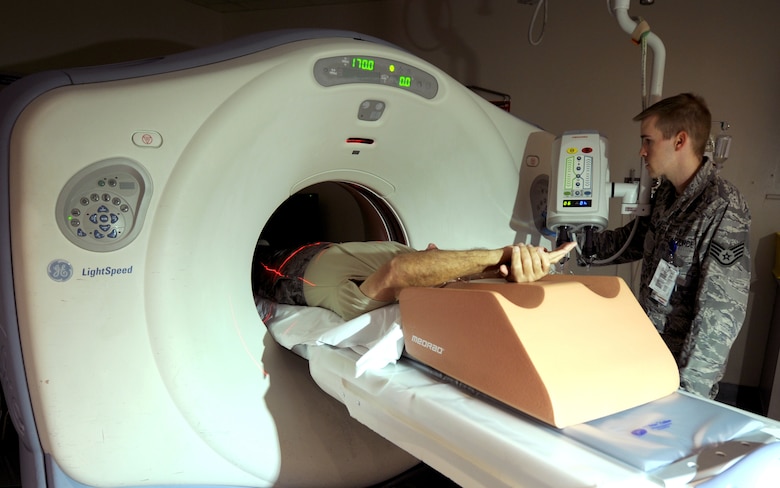
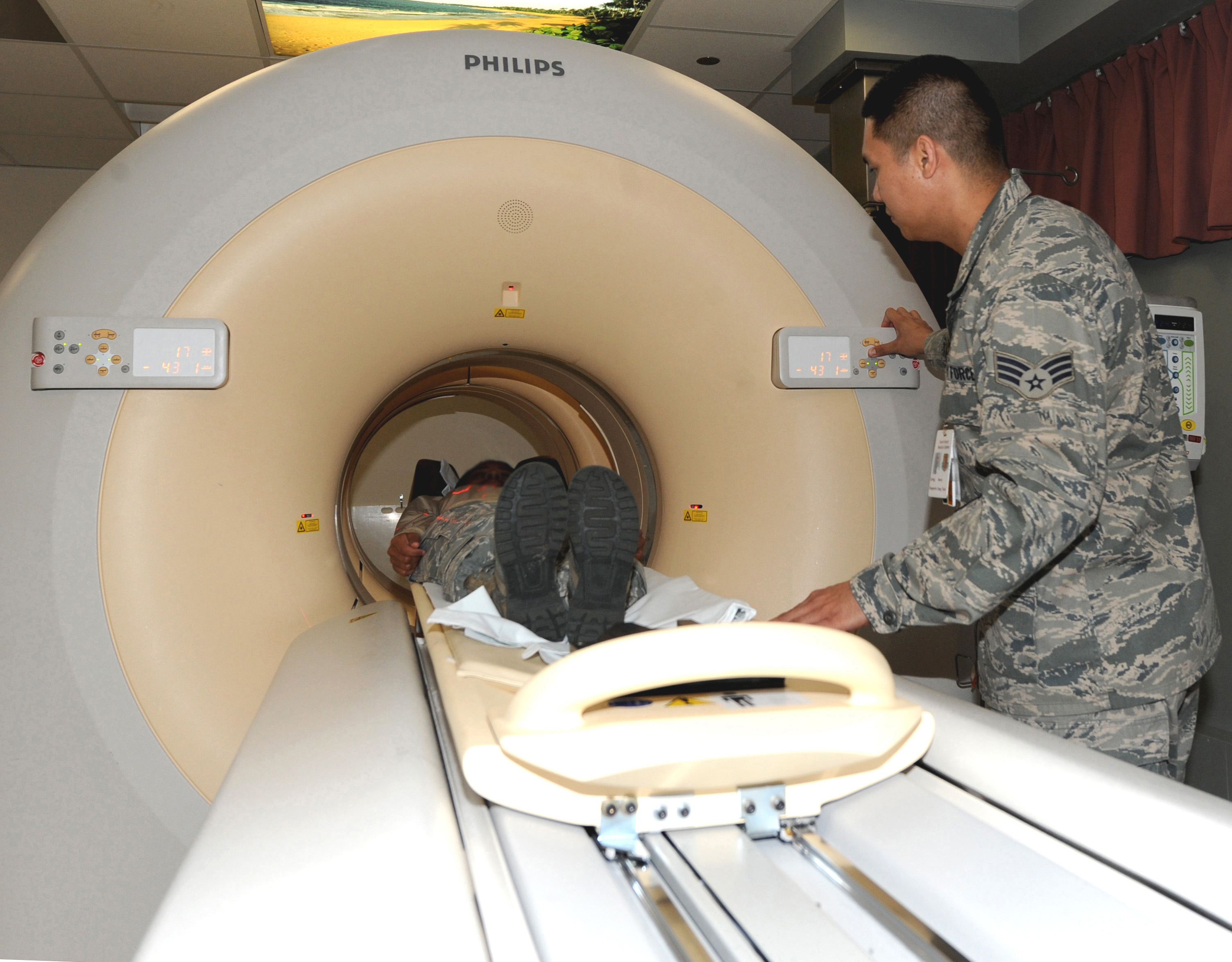
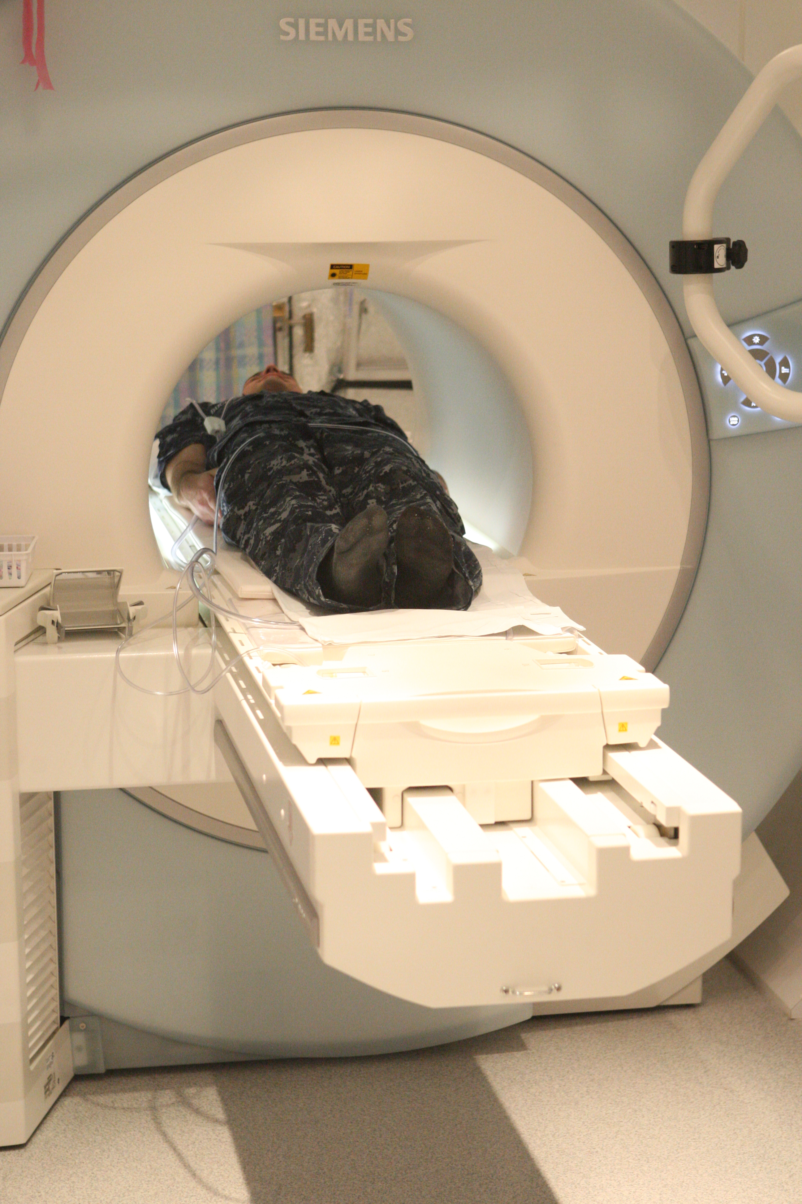
While positioning you on the examination table, the technologist will explain your treatment and respond to any concerns you might have. If contrast color is being utilized, it will be injected through your IV. Throughout the injection, you might experience a warm experience all over your body and a metal taste in your mouth. This is typical. If you experience any itching, sneezing, nasal blockage, scratchy throat or swelling of your face, please notify the technologist right away.

Most often, you will be asked to lie flat on your back with your arms over your head. Some scans may need you to be positioned in your corner or stomach. The table you are on will slide into the scanner. The scanner is open at the back and the front, enabling you to see out. The technologist will always be able to see and hear you throughout your examination. You will be asked to hold extremely still and at times to hold your breath. This treatment usually takes between 15 and 30 minutes.


A CT (computed tomography) scan uses X-rays to make in-depth pictures of your body and structures inside your body. A CT scan of the chest can give your medical professional details about your lungs, your heart, and other structures in your chest. Throughout the test, you will rest on a table that is connected to the CT scanner. The CT scanner is a large doughnut-shaped maker.

UBid Imaging and Diagnostics, LLC

Business Office

Tollfree 855-286-7400

2135 City Gate Lane,

Suite 300

Naperville, IL 60563


Posted by edwinmqai906 at 10:11 AM EDT
Post Comment | Permalink | Share This Post
Monday, 25 March 2019
Ct Scan: Expectations vs. Reality

You will be asked to push a narrow table that moves into the center of the scanner. As soon as you are inside the scanner, the machine's x-ray beam turns around you. Little detectors inside the scanner measure a variety of x-rays that make it through the part of the body being studied. A computer system takes this details and uses it to develop numerous private images, called slices. These images can be kept, seen on a monitor, or printed on film. Three-dimensional images of organs can be produced by stacking the private pieces together.

You need to be still during the examination due to the fact that movement triggers blurred images. You may be informed to hold your breath for short time periods. Normally, complete scans take only a few minutes. The latest multidetector scanners can image your whole body in less than 30 seconds. Certain CT scans require an unique color, called contrast, to be delivered into the body prior to the test starts. Contrast can highlight specific locations inside the body, which creates a clearer image. If your medical professional requests a CT scan with contrast, you will be given it through a vein (IV) in your arm or hand.

CT scans and other x-rays are strictly kept an eye on and managed to make certain https://www.washingtonpost.com/newssearch/?query=CT Scan they utilize the least amount of radiation. CT scans use low levels of ionizing radiation, which has the potential to trigger cancer and other defects. Nevertheless, the threat from any one scan is little. The risk increases as many more studies are done. Sometimes, a CT scan may still be done if the benefits greatly surpass the threats. For example, it can be riskier to not have the test if your physician thinks you may have cancer.

The most typical type of contrast given into a vein includes iodine. If an individual with an iodine allergy is provided this type of contrast, queasiness, sneezing, throwing up, itching, or hives might occur. In unusual cases, the color can cause a life-threatening allergic response called anaphylaxis. If you have any difficulty breathing throughout the test, you should notify the scanner operator right away. Scanners include an intercom and speakers, so the operator can hear you at all times. In individuals with kidney issues, the color might have hazardous impacts on the kidneys. In these scenarios, unique steps may be taken to make the contrast color safer to utilize.

Depending upon the reason for your examination you may have an I.V. placed in your arm. Through this I.V. a contrast will be injected. During the examination, you will push a table with your arms raised above your head. The table will rise and move in and out of a big circle once the examination has begun. The technologist more info will describe any breathing directions you might have during the real test and cover some particular medical history concerns.

After this is complete the scan will begin. A few images (photos) will be taken so that the technologist can set up the area to be scanned. At this point, it will be very important that you hold still. If you are receiving an injection of contrast, a technologist will stay in the space for the start of the injection and march just prior to the scan starting once again. The majority of the time clients will have this test done lying on their back however there are some clients that will require to rest on their stomach.

UBid Imaging and Diagnostics, LLC

Business Office

Tollfree 855-286-7400

2135 City Gate Lane,

Suite 300


Naperville, IL 60563


Posted by edwinmqai906 at 2:51 PM EDT
Post Comment | Permalink | Share This Post
Friday, 15 March 2019
Benefits Of CT Scan That May Adjustment Your Viewpoint

Chest CT scans are more diagnostically precise than chest radiography for postmortem rib fracture detection in cases of physical abuse in kids no matter a radiologist's experience or fracture location, according to research study published in the Sept. concern of The Lancet.

Rib fractures in babies and kids are extremely particular to physical abuse. In 30 percent of abuse cases, rib fractures are the only skeletal irregularity present and are discovered in a third of infants who die due to physical abuse, wrote lead author Own Arthurs, PhD, an expert pediatric radiologist at the Great Ormond Street Hospital for Children in London, and associates.

Although chest radiography is the basic technique for identifying rib fractures, the scientists explained chest CT Contrast and multiple scans could provide greater accuracy and supply clearness and assistance for medicolegal procedures in physical abuse cases.

Data from the radiology information system at the Great Ormond Street Healthcare facility was analyzed for all children aged zero to 16 years who had a full-body radiography exam, CT scan and full autopsy between January 2012 through January 2017 for forensic investigation.


An overall of 25 cases of children (between the ages of one month to 7 years) with a combined total of 136 rib fractures were picked for analysis.

An overall of 38 global volunteer radiologists differing in level of experience were blinded to the clinical image details. All analyzed the set of chest radiographs to identify the presence of rib fractures, fracture location and reported the confidence level of their analysis. One month later on, the radiologists compared their detection of rib fractures in the chest radiographs to CT scans of the exact same cases.

In general, all 38 radiologists discovered three-times as lots of rib fractures properly identified by chest CTs compared to chest radiography, the researchers wrote. Chest CT also exceeded radiography in detection level of sensitivity of the proper rib and diagnosis of a patient with any rib fracture. All radiologists reported to have higher confidence when utilizing CT imagies than radiographs, also.

The scientists advise the use of chest CT in both post-mortem assessment of suspected children physical abuse cases along with in cases in living children when https://www.washingtonpost.com/newssearch/?query=CT Scan chest radiography is unfavorable or unclear.

" Together with proof that health centers, social workers, moms and dads, and kids adhere badly to the schedule of follow-up radiographs, we argue that using CT in the severe setting in which radiographs are negative or indisputable will most likely determine otherwise undiscovered rib fractures, offering instant diagnostic info, which is preferable to waiting for follow-up radiographs," Arthurs et al. composed. "Because all infants presumed of physical abuse will go through a head CT to try to find intracranial injury based on national guidelines, we argue that the addition of a subsievert chest CT throughout the same presence would be pragmatic."

UBid Imaging and Diagnostics, LLC

Business Office

Tollfree 855-286-7400

2135 City Gate Lane,

Suite 300

Naperville, IL 60563


Posted by edwinmqai906 at 7:49 PM EDT
Post Comment | Permalink | Share This Post
Wednesday, 13 March 2019
Typical Misconceptions About CT Scan

A chest CT scan http://edition.cnn.com/search/?text=CT Scan is a more in-depth kind of chest x-ray. This painless imaging test takes numerous comprehensive pictures, called pieces, of your lungs and the inside of your chest. Computers can combine these images to produce three-dimensional (3D) designs to assist reveal the size, shape, and position of your lungs and structures in your chest. This imaging test is often done to act on abnormal findings from earlier chest x rays. A chest CT scan also can assist identify the cause of lung symptoms such as shortness of breath or chest pain, or inspect to see if you have particular lung issues such as a growth, excess fluid around the lungs that is referred to as pleural effusion, pulmonary embolism, emphysema, tuberculosis, and pneumonia.

Your chest CT scan may be carried out in a medical imaging center or medical facility. The CT scanner is a big, tunnel-like machine that has a table. You will lie still on the table and the table will move into the scanner. Talk with your doctor if you are uneasy in tight or closed areas to see if you need medication to relax you throughout the test. You will hear soft buzzing or clicking sounds when you are inside the scanner and the scanner is taking images. You will have the ability to speak with and speak to the professional carrying out the test while you are inside the scanner. For some medical diagnoses, a contrast dye, often iodine-based, may be injected into a vein in your arm before the imaging test. This contrast dye highlights locations inside your chest and produces clearer photos. You may feel some pain from the needle or, after the contrast color is injected, you may feel warm briefly or have a short-lived metal taste in your mouth.


Chest CT scans have some risks. In rare circumstances, some people have an allergy to the contrast color. There is a small threat of cancer, particularly in growing kids, because the test utilizes radiation. Although the amount of radiation from one test is usually less than the quantity of radiation you are naturally exposed to over 3 years, patients must not receive more CT scans than the number that clinical standards recommend. Another danger is that chest CT scans might discover an incidental finding, which is something that doesn't trigger symptoms today might need more tests after being discovered. Speak to your physician and the service technicians carrying out the test about whether you are or could be pregnant. If the test is not urgent, they may have you wait to do the test till after your pregnancy. If it is urgent, the specialists will take additional actions to protect your infant throughout this test. Let your physician understand if you are breastfeeding because contrast color can pass into your breast milk. If you need to have a contrast color injected, you may want to pump and conserve enough breast milk for one to 2 days after your test or you might bottle-feed your baby for that time.

Calculated tomography (CT scan or FELINE scan) is a noninvasive diagnostic imaging procedure that uses a combination of X-rays and computer system innovation to produce horizontal, or axial, images (frequently called pieces) of the body. A CT scan programs detailed pictures of any part of the body, including the bones, muscles, fat, and organs. CT scans are more comprehensive than basic X-rays.

In standard X-rays, a beam of energy is aimed at the body part being studied. A plate behind the body part records the variations of the energy beam after it passes through skin, bone, muscle, and other tissue. While much details can be obtained from a standard X-ray, a lot of information about internal organs and other structures is not available. CT Scan Naperville IL UBid Imaging In computed tomography, the X-ray beam moves in a circle around the body. This permits several views of the same organ or structure. The X-ray info is sent to a computer system that translates the X-ray data and shows it in a two-dimensional (2D) type on a screen.

UBid Imaging and Diagnostics, LLC

Business Office

Tollfree 855-286-7400

2135 City Gate Lane,

Suite 300

Naperville, IL 60563


Posted by edwinmqai906 at 10:02 AM EDT
Post Comment | Permalink | Share This Post
Friday, 22 February 2019
Facts You Never Knew About CT Scan

Calculated tomography (CT) of the chest utilizes special x-ray equipment to examine irregularities found in other imaging tests and to help identify the cause of an inexplicable cough, shortness of breath, chest discomfort, or fever. CT scanning is quickly, painless, noninvasive and accurate. Since it has the ability to detect extremely small blemishes in the lung, chest CT is particularly efficient for detecting lung cancer at its earliest, most treatable stage.

Tell your medical professional if there's a possibility you are pregnant and go over any recent health problems, medical conditions, medications you're taking, and allergic reactions. You will be advised not to eat or consume anything for a few hours ahead of time. If you have a known allergy to contrast product, your medical professional might prescribe medications to reduce the danger of an allergy. These medications need to be taken 12 hours prior to your test. Leave fashion jewelry in the house and wear loose, comfortable clothing. You might be asked to use a gown.

CT scans and other x-rays are strictly monitored and controlled to ensure they utilize the least amount of radiation. CT scans use low levels of ionizing radiation, which has the prospective to cause cancer and other defects. Nevertheless, the threat from any one scan is little. The danger increases read more as a lot more studies are done. In some cases, a CT scan might still be done if the advantages significantly surpass the risks. For example, it can be riskier to not have the exam if your medical professional believes you might have cancer.

The most typical kind of contrast given into a vein consists of iodine. If an individual with an iodine allergy is given this kind of contrast, nausea, sneezing, throwing up, itching, or hives might happen. In unusual cases, the dye can cause a lethal allergic response called http://query.nytimes.com/search/sitesearch/?action=click&contentCollection®ion=TopBar&WT.nav=searchWidget&module=SearchSubmit&pgtype=Homepage#/CT Scan anaphylaxis. If you have any difficulty breathing during the test, you should inform the scanner operator right away. Scanners include an intercom and speakers, so the operator can hear you at all times. In individuals with kidney problems, the color may have damaging effects on the kidneys. In these scenarios, special actions may be taken to make the contrast color much safer to use.

Computed tomography, more commonly referred to as a CT or CAT scan, is a diagnostic medical test that, like standard x-rays, produces several images or images of the inside of the body. The cross-sectional images generated throughout a CT scan can be reformatted in multiple aircrafts, and can even generate three-dimensional images. These images can be viewed on a computer system display, printed on movie or moved to a CD or DVD. CT pictures of internal organs, bones, soft tissue and blood vessels typically offer higher detail than traditional x-rays, particularly of soft tissues and blood vessels.

Using a variety of techniques, including adjusting the radiation dosage based upon client size and new software application innovation, the quantity of radiation needed to carry out a chest CT scan can be considerably reduced. A low-dose chest CT produces pictures of enough image quality to spot many lung illness and irregularities utilizing substantially less ionizing radiation than a traditional chest CT scan-- in some cases lowering dose by 65 percent or more. Low dosage chest CT is routinely used for evaluation of obtained and congenital lung irregularities, such as pneumonia, interstitial lung illness or growth assessment. There is continuous research to lower radiation dosages even further. Your radiologist will decide the appropriate settings to be used for your scan depending upon your medical issues and what info is required from the CT scan. If your kid is to have a CT scan, the appropriate low-dose pediatric settings should be used.

UBid Imaging and Diagnostics, LLC

Business Office

Tollfree 855-286-7400

2135 City Gate Lane,

Suite 300


Naperville, IL 60563


Posted by edwinmqai906 at 8:33 AM EST
Post Comment | Permalink | Share This Post
Monday, 11 February 2019
Little Understood Ways to CT Scan of Brain

A CAT scan of the head (likewise called a head CT) is a pain-free test that uses an unique X-ray device to take images of a client's brain, skull, and sinuses, in addition to capillary in the head. The doughnut-shaped device circles the head, taking pictures to offer cross-sections of the brain from different angles. These photos are sent out to a computer that records the images. It also can put them together to form three-dimensional images. A radiology technician carries out the CAT scan (likewise called a CT scan or a computed axial tomography scan).

A CT brain is bought to look at the structures of the brain and examine for the existence of pathology, such as mass/tumor, fluid collection (such as an abscess), ischemic processes (such as a stroke). It is particularly helpful for hemorrhage, trauma or fracture to the skull and for hydrocephalus.

The CT head scan is a computer-generated series of images from numerous X-rays taken at various levels. Great X-ray beams passed through the topic are taken in to various degrees by different tissues and the transmitted radiation is determined by a scanning gadget. The degree of absorption of X-rays is proportional to the density of the tissue through which it passes. Hounsfield units (HU) are used to measure just how much of the X-ray beam is soaked up by the tissues at each point in the body. The denser the tissue, the more the X-ray beam is attenuated and the greater the number. Systems are developed on a relative scale with water as the recommendation point. Water is constantly 0HU, bone is approximately 1000HU and air is-- 1000HU.

The CAT https://www.washingtonpost.com/newssearch/?query=MRI Scans Of The Head scan (also called CT scan) is widely known by name, but do you actually understand what it is and comprehend how it works? A CT scan is normally one of the first tests performed in a stroke evaluation, especially during an intense stroke in the emergency clinic. This test can show areas of problems in the brain and can assist to figure out if these areas are caused by inadequate blood circulation (ischemic stroke), a burst blood vessel (hemorrhage), or a various type of an issue. CT scans can be acquired on any part of the body, but the details here applies only to CT scans of the head. A CT scan usages X-rays to take pictures of your skull and brain. The patient lies in a tunnel-like maker while the within the machine turns and takes X-rays of the head from various angles. These pictures are later used by computers to make a picture of a "slice" (or cross-section) of the brain.

Computed tomography (CT) of the head or Computed Axial Tomography (CAT) scanning uses a series of x-rays of the head drawn from various instructions. Normally utilized for rapidly viewing brain injuries, CT scanning uses a computer program that carries out a numerical integral computation (the inverse Radon transform) on the determined x-ray series to approximate how much Check out here of an x-ray beam is absorbed in a little volume of the brain. Normally the details is presented as a series of cross-sections of the brain.


UBid Imaging and Diagnostics, LLC

Business Office

Tollfree 855-286-7400

2135 City Gate Lane,

Suite 300

Naperville, IL 60563


Posted by edwinmqai906 at 2:07 PM EST
Post Comment | Permalink | Share This Post
Sunday, 3 February 2019
Here Is What You Should Provide for Your CT Scan of Brain

Computed tomography (CT or FELINE scan) is a noninvasive diagnostic imaging procedure that uses a combination of X-rays and computer system innovation to produce horizontal, or axial, images (often called pieces) of the body. A CT scan programs detailed pictures of any part of the body, consisting of the bones, muscles, fat, and organs. CT scans are more in-depth than standard X-rays. In standard X-rays, a beam of energy is focused on the body part being studied. A plate behind the body part catches the variations https://ubidmri.com of the energy beam after it passes through skin, bone, muscle, and other tissue. While much details can be gotten from a basic X-ray, a lot of detail about internal organs and other structures is not readily available.

In computed tomography, the X-ray beam relocations in a circle around the body. This enables several views of the very same organ or structure. The X-ray info is sent to a computer that interprets the X-ray information and displays it in a two-dimensional (2D) form on a monitor. CT scans may be done with or without "contrast." Contrast refers to a compound taken by mouth or injected into an intravenous (IV) line that causes the specific organ or tissue under research study to be seen more clearly. Contrast assessments might need you to quick for a certain amount of time before the treatment. Your physician will notify you of this prior to the treatment. CT scans of the brain can provide more comprehensive info about brain tissue and brain structures than basic X-rays of the head, thus supplying more info associated to injuries and/or diseases of the brain.

A CT scan uses X-rays and computers to make images of the body. It can sometimes assist doctors diagnose headaches and their causes. You might need one if you have headaches day-to-day or almost every day or have an unexpected onset extreme headache. Medical professionals can't identify migraines with the test, though. The test is pain-free. To get the scan, you'll push a table. You may get a shot of "contrast product" into one of your veins, which will help doctors see parts of your brain more clearly on the image. Be sure to tell the physician or nurse if you have actually had an allergy to contrast material in the past. Your doctor will likewise need to check your kidney function prior to using contrast. The dyes have iodine, which can cause a reaction in some people.

A computed tomography (CT) scan usages X-rays to make images of the head and face. During the test, you will rest on a table that is attached to the CT scanner, which is a large doughnut-shaped machine. Your head will be positioned inside the scanner. The CT scanner sends out X-rays through the head. Each rotation of the scanner supplies an image of a thin piece of the head and face. One part of the scanning maker can tilt to take images from various positions. All of the photos are conserved as a group on a computer. They also can be printed.

Sometimes, a dye called contrast product might be put in a vein (IV) in your arm or into the back canal. The color makes structures and organs much easier to see https://www.washingtonpost.com/newssearch/?query=MRI Scans Of The Head on the CT images. The color might be used to check blood flow and try to find tumors, areas of swelling, or nerve damage.


UBid Imaging and Diagnostics, LLC

Business Office

Tollfree 855-286-7400

2135 City Gate Lane,

Suite 300

Naperville, IL 60563


Posted by edwinmqai906 at 11:20 AM EST
Post Comment | Permalink | Share This Post
Thursday, 24 January 2019
The Unknown Secret To Mastering CT Scan of Brain

Calculated tomography (CT) of the head utilizes special x-ray devices to help examine head injuries, extreme headaches, dizziness, and other symptoms of an aneurysm, bleeding, stroke and brain growths. It likewise assists your medical professional to examine your face, sinuses, and skull or to prepare radiation therapy for brain cancer. In emergency situation cases, it can expose internal injuries and bleed quickly enough to assist in saving lives.

Inform your medical professional if there's a possibility you are pregnant and talk about any recent illnesses, medical conditions, medications you're taking, and allergies. You will be instructed not to eat or consume anything for a few hours ahead of time. If you have a known allergy to contrast product, your doctor may recommend medications to reduce the danger of an allergic reaction. These medications need to be taken 12 hours prior to your test. Leave jewelry in the house and use loose, comfortable clothes. You might be asked to wear a dress.


Computed tomography, more typically called a CT or FELINE scan, is a diagnostic medical test that, like traditional x-rays, produces multiple images or http://query.nytimes.com/search/sitesearch/?action=click&contentCollection®ion=TopBar&WT.nav=searchWidget&module=SearchSubmit&pgtype=Homepage#/MRI Scans Of The Head pictures of the within the body. The cross-sectional images produced throughout a CT scan can be reformatted in numerous airplanes, and can even produce three-dimensional images. These images can be seen on a computer screen, printed on film or transferred to a CD or DVD. CT images of internal organs, bones, soft tissue and capillary usually offer greater detail than standard x-rays, especially of soft tissues and capillary. CT scanning offers more in-depth information on head injuries, stroke, brain growths and other brain diseases than regular radiographs (x-rays).

Metal things, including fashion jewelry, glasses, dentures and barrettes, may affect the CT images and need to be left at home or eliminated prior to your test. You may also be asked to eliminate hearing aids and detachable dental work. Females will be asked to remove bras containing metal underwire. You might be asked to remove any piercings, if possible.

You will be asked not to consume or drink anything for a few hours ahead of time, as contrast product will be used in your test. You need to inform your physician of all medications you are taking and if you have any allergies. If you have a known allergic reaction to contrast product, or "color," your doctor might prescribe medications (usually a steroid) to reduce the risk of an allergic reaction. These medications typically require to be taken 12 hours prior to administration of contrast material. To avoid unneeded hold-ups, call your doctor prior to the exact time of your examination.

Likewise, notify your doctor of any current health problems or other medical conditions and whether you have a history of heart problem, asthma, diabetes, kidney disease or thyroid issues. Any of these conditions might increase the risk of an uncommon adverse result. The radiologist likewise must know if you have asthma, numerous myeloma or any disorder of the heart, kidneys or thyroid gland, or if you have diabetes-- especially if you are taking Glucophage. Women must constantly inform their doctor and the CT technologist if there is any possibility that they might https://ubidmri.com be pregnant. See the Security page to find out more about pregnancy and x-rays.

UBid Imaging and Diagnostics, LLC

Business Office

Tollfree 855-286-7400

2135 City Gate Lane,

Suite 300

Naperville, IL 60563


Posted by edwinmqai906 at 11:43 AM EST
Post Comment | Permalink | Share This Post

Newer | Latest | Older