

Computed tomography (CT or FELINE scan) is a noninvasive diagnostic imaging procedure that uses a combination of X-rays and computer system innovation to produce horizontal, or axial, images (often called pieces) of the body. A CT scan programs detailed pictures of any part of the body, consisting of the bones, muscles, fat, and organs. CT scans are more in-depth than standard X-rays. In standard X-rays, a beam of energy is focused on the body part being studied. A plate behind the body part catches the variations https://ubidmri.com of the energy beam after it passes through skin, bone, muscle, and other tissue. While much details can be gotten from a basic X-ray, a lot of detail about internal organs and other structures is not readily available.
In computed tomography, the X-ray beam relocations in a circle around the body. This enables several views of the very same organ or structure. The X-ray info is sent to a computer that interprets the X-ray information and displays it in a two-dimensional (2D) form on a monitor. CT scans may be done with or without "contrast." Contrast refers to a compound taken by mouth or injected into an intravenous (IV) line that causes the specific organ or tissue under research study to be seen more clearly. Contrast assessments might need you to quick for a certain amount of time before the treatment. Your physician will notify you of this prior to the treatment. CT scans of the brain can provide more comprehensive info about brain tissue and brain structures than basic X-rays of the head, thus supplying more info associated to injuries and/or diseases of the brain.
A CT scan uses X-rays and computers to make images of the body. It can sometimes assist doctors diagnose headaches and their causes. You might need one if you have headaches day-to-day or almost every day or have an unexpected onset extreme headache. Medical professionals can't identify migraines with the test, though. The test is pain-free. To get the scan, you'll push a table. You may get a shot of "contrast product" into one of your veins, which will help doctors see parts of your brain more clearly on the image. Be sure to tell the physician or nurse if you have actually had an allergy to contrast material in the past. Your doctor will likewise need to check your kidney function prior to using contrast. The dyes have iodine, which can cause a reaction in some people.
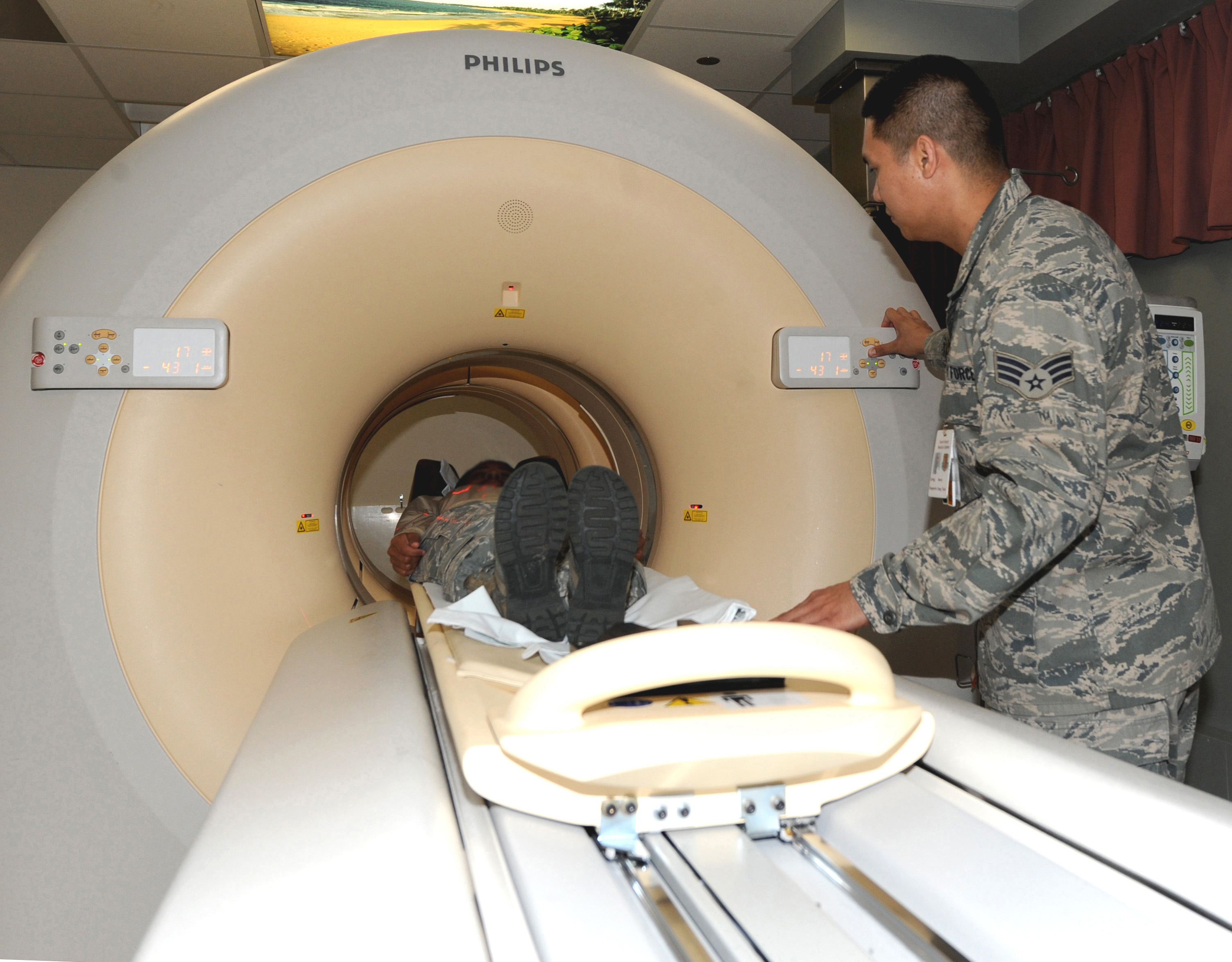
A computed tomography (CT) scan usages X-rays to make images of the head and face. During the test, you will rest on a table that is attached to the CT scanner, which is a large doughnut-shaped machine. Your head will be positioned inside the scanner. The CT scanner sends out X-rays through the head. Each rotation of the scanner supplies an image of a thin piece of the head and face. One part of the scanning maker can tilt to take images from various positions. All of the photos are conserved as a group on a computer. They also can be printed.
Sometimes, a dye called contrast product might be put in a vein (IV) in your arm or into the back canal. The color makes structures and organs much easier to see https://www.washingtonpost.com/newssearch/?query=MRI Scans Of The Head on the CT images. The color might be used to check blood flow and try to find tumors, areas of swelling, or nerve damage.
UBid Imaging and Diagnostics, LLC
Business Office
Tollfree 855-286-7400
2135 City Gate Lane,
Suite 300
Naperville, IL 60563